MRI Safety Testing for Medical Devices
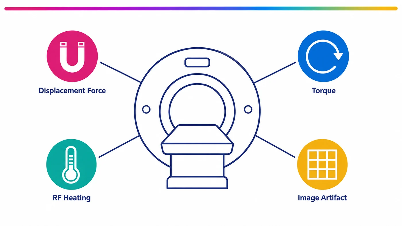
Biotech Anatomy evaluates how a medical device behaves inside a 3 Tesla magnetic field. The work is carried out at research and development level, on a whole body clinical scanner, for devices that are still in development, with your engineering team present and able to change the test while it runs. This page sets out what the evaluation measures, which tests are running today, which are still being built, and where this stage sits in relation to an accredited regulatory campaign.
Reading time: 8 minutes
Key points
- Testing runs on a 3T MAGNETOM Vida whole body scanner with a 70 cm wide bore, operating system XA60.
- Image artifact and displacement force evaluations run today. Torque and radiofrequency heating methods for non active implants are in development.
- The results are generated at a research‑and‑development stage and therefore do not yet constitute accredited certification data nor are they intended for regulatory submission at this time; nonetheless, they provide supportive evidence for prototype‑level evaluation with respect to MRI compatibility.
- Your team is invited into the session and can adjust device orientation, configuration and scan parameters while the scanner is running.
Contents
- The four physical effects an MRI environment produces
- The 3T scanner and its 70 cm bore
- Tests running now and tests in development
- Where this stage sits before accreditation
- What the laboratory needs beforehand
- Safety screening for everyone entering the magnet room
- Running MRI evaluation alongside cadaveric pre-clinical work
The four physical effects an MRI environment produces
An MRI scanner exposes a device to three separate fields at once, and each acts on the device differently.
The static magnetic field is always on. It pulls ferromagnetic material toward the bore, and it twists any object whose magnetic properties vary along its length. Those are two distinct effects and they are measured separately, as displacement force and as torque.
The radiofrequency field is transmitted in pulses. Conductive structures inside a device can absorb that energy and convert it to heat at the device surface, which matters most for elongated conductive geometry.
The device also distorts the field it sits in. That distortion appears in the image as signal loss, signal pile up or geometric shift around the device, and it determines whether a clinician can still read the anatomy next to the implant.
The published methods that define how each effect is measured
Four ASTM test methods define the measurement for each effect. ASTM F2052 covers magnetically induced displacement force, ASTM F2213 covers magnetically induced torque, ASTM F2182 covers radiofrequency induced heating, and ASTM F2119 covers evaluation of MR image artifacts.
Naming a method describes what is being measured and how it is measured. That is a separate matter from holding accreditation to certify a device against it, and the difference is set out below.
Those measurements exist to support a labelling decision. ASTM F2503 defines how a device is marked for the MR environment, as MR Safe, MR Conditional or MR Unsafe. MR Conditional is what most development programmes are working toward, and the label carries the conditions the device was shown to be safe under, field strength among them. Which classification you are targeting decides which effects matter most at this stage.
The 3T scanner and its 70 cm bore
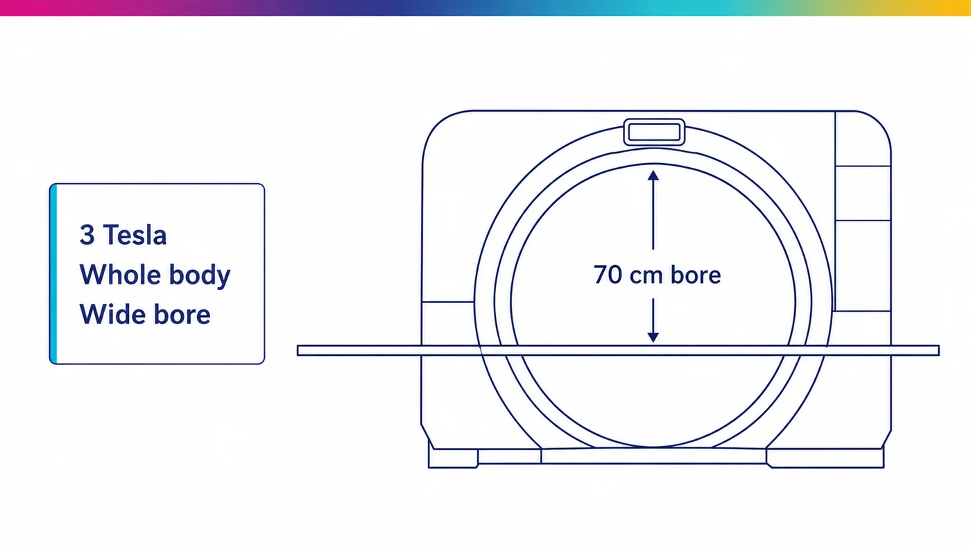
Testing runs on a MAGNETOM Vida operating at 3 Tesla, with system software XA60. It is a whole body scanner with a 70 cm wide bore.
Two properties of that configuration matter when you plan a test. Field strength sets the magnitude of every static field effect, because displacement force and torque both scale with the static field and its spatial gradient. A device that behaves acceptably at 1.5T can behave differently at 3T. Most MR Conditional labelling covers both, so a device intended for both needs evidence at both. Say which field strengths your labelling will claim before the session is planned.
Bore diameter sets what will physically fit. A 70 cm bore accepts test fixtures, phantom containers and positioning apparatus that a narrower bore will not, which matters when a device has to be held at a defined orientation and distance rather than simply laid on the table.
Tests running now and tests in development
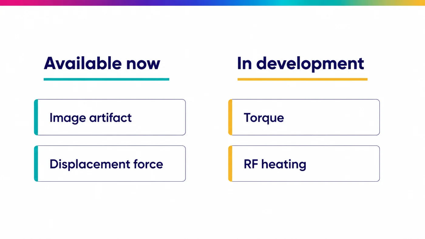
The division is new and the test menu is being built in stages. The position is set out plainly below, because a method that is still being developed cannot be booked.
| Evaluation | Reference method | Status |
|---|---|---|
| Image artifact | ASTM F2119 | Running |
| Displacement force | ASTM F2052 | Running |
| Magnetically induced torque | ASTM F2213 | In development |
| Radiofrequency induced heating | ASTM F2182 | In development |
Torque and heating are being developed for non active implants, meaning implants containing no electrical circuit and no power source.
Active implantable devices, which include pacemakers, neurostimulators and anything carrying its own power source, are assessed under ISO/TS 10974. That specification adds gradient induced voltage, device function during exposure and numerical modelling to the physical measurements above, and it is not in scope for this service.
If your program needs a method currently listed as in development, say so anyway. Which methods are being asked for is what decides the order they are completed in.
Where this stage sits before accreditation
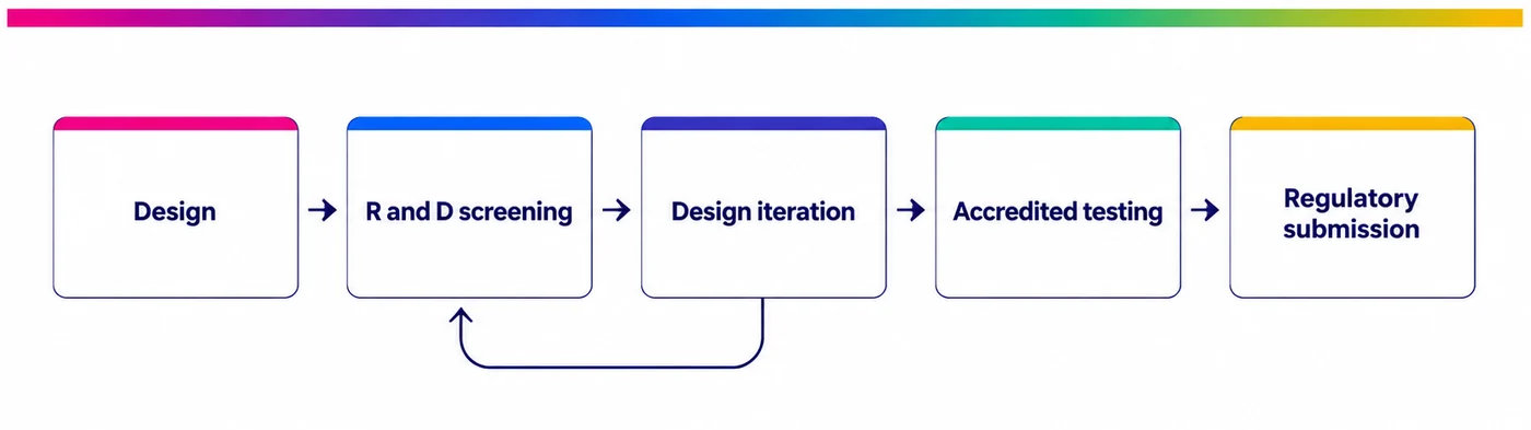
The results this laboratory produces (for now) are research and development results. They are not-yet accredited certification results, and they are not intended for submission to the FDA or to a European notified body. Regulatory submission requires a laboratory holding ISO/IEC 17025 accreditation for the specific method, and the FDA guidance on testing and labelling devices for the MR environment sets out what a submission is expected to contain.
That is a real limit, and it is stated plainly because the alternative wastes your time. It is also not what this stage is for.
A device that displaces at 3T displaces regardless of who measures it. Finding that out in an R&D session costs one session. Finding it out partway through an accredited campaign costs the campaign, the schedule, and every design decision built on top of it.
This stage is built for:
- screening candidate materials and configurations before committing to one
- comparing design variants against each other under identical conditions
- generating internal evidence for a design review or a risk file
- establishing which methods and which orientations to book at the accredited laboratory
- resolving whether a suspected problem is real before it is escalated
It is not, yet, built for regulatory submission, for an MR Conditional labelling claim, or as evidence in a CE or FDA file.
Not sure which effects your device needs measured first
Send the device specification and we will tell you what is worth measuring at this stage and what belongs in an accredited campaign.
What the laboratory needs beforehand
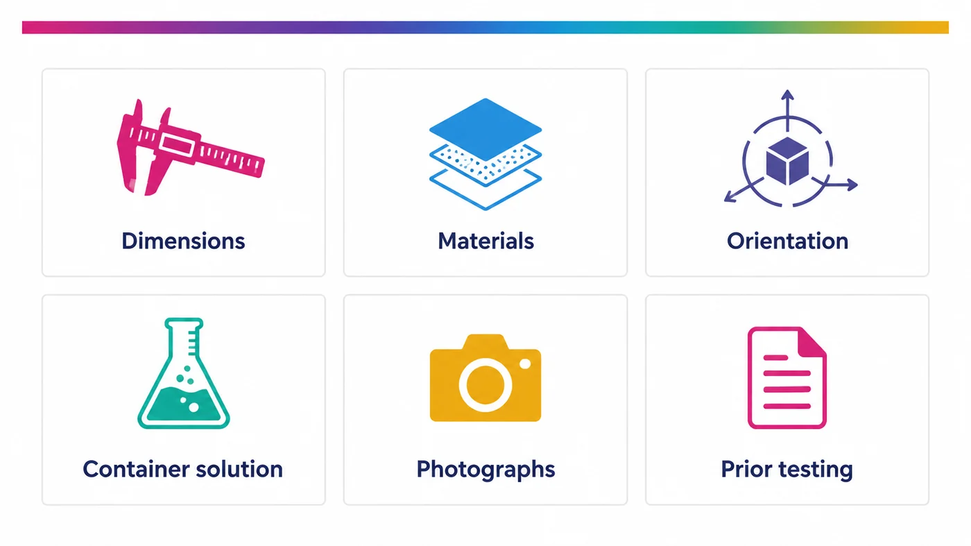
Preparation depends almost entirely on information supplied in advance, because the container, the fixture and the scan protocol are all built around the specific device. The intake covers:
- A full list of materials in the device, with quantities.
- Whether the device contains an electrical circuit requiring a power source.
- Dimensions: length, width, depth and total volume.
- Configuration, including whether the device is modular and supports more than one assembly.
- The organ the device is used in, and its clinical purpose.
- The likely orientation inside the body: parallel to the long axis, left to right, front to back, or variable.
- Any previous MRI assessment, including which effects were measured and at what field strength.
- Which container solutions will not damage the device. The standard options are copper sulphate, nickel chloride and manganese chloride.
- Photographs of the device from several angles, with a scale in frame.
Two requests make more difference than the rest combined.
Send the worst case sample.
That means the largest variant, the fullest configuration, the version most likely to fail. Testing a benign variant tells you very little about the one that will actually be marketed, and it is the marketed one that has to carry the labelling.
Send a physical sample in advance where you can. A unit destined for the bin, a used unit or a malfunctioning unit is enough. It lets the container and the fixture be adapted before the session rather than during it, which is where scanner time is normally lost.
Safety screening for everyone entering the magnet room
The static field is always on at 3 Tesla, roughly twice the strength of a scrapyard lifting magnet. It does not affect biological tissue, but it acts strongly on metal and it will corrupt electronic circuits and magnetically stored data.
Everyone attending a session completes a safety questionnaire in advance. It covers implants, any medical device attached to the body, and whether all metal and devices can be removed before entry. This applies to your engineers and to observers exactly as it applies to laboratory staff.
Device information supplied for a test is treated as confidential.
It is used only for that scan, and disclosed only to the personnel directly involved in it, all of whom are covered by signed institutional non disclosure agreements.
Running MRI evaluation alongside cadaveric pre-clinical work
Biotech Anatomy is the only private Israeli company dedicated to employing human cadaver for research. A device program needing both a human anatomical model and MRI evaluation can plan both through one relationship, with a team that already holds the device specification, the orientation data and the handling requirements from the first study. Our cadaver laboratory and this service run from the same program team.
Where the anatomical fit question and the MRI safety question interact, that continuity is worth more than it sounds. The orientation a device actually adopts once placed is an input to the displacement and torque assessment, and it is usually an assumption. A program that has already run the placement work has the benefit of actual knowledge. The same applies across our wider medical device testing work.
Specimen selection, provenance and documentation for that side of the work are covered under procuring human tissue specimens.
Frequently asked questions
Can our engineers attend the test?
Yes. Clients are invited to be present and involved in how the test is carried out. Orientation, configuration and scan parameters can be adjusted during the session, which is one of the main reasons to run this stage in a research and development setting rather than a fixed protocol one.
Can we use the results in an FDA or CE submission?
Not yet. Results are produced at research and development level, not certification level. Regulatory submission requires a laboratory holding ISO/IEC 17025 accreditation for the specific method. This stage is what you do before that, to decide what to send and to avoid discovering a problem inside an accredited campaign.
Do you test active implantable devices?
Not currently. Development is focused on non active implants, meaning devices with no electrical circuit and no power source. Active implantable devices are assessed under ISO/TS 10974, which adds gradient induced voltage, device function during exposure and numerical modelling to the physical measurements.
Which field strength do you test at?
Three Tesla, on a whole body MAGNETOM Vida with a 70 cm bore. If your labelling will also claim 1.5T, say so when the test is planned. Static field effects scale with field strength, so evidence at one strength does not carry across to the other.
What should we send before the session?
Materials and quantities, dimensions, configuration, intended orientation in the body, any previous MRI assessment, and which container solutions are safe for the device, together with photographs from several angles. Where possible send a physical sample in advance, including a used or non functional unit, so the container and fixture can be prepared before scanner time starts.
Planning MRI evaluation for a device in development
Tell us what the device is, where it sits in the body, and what your labelling will claim. We will set out what a session can and cannot establish.
About Biotech Anatomy

Biotech Anatomy LTD has provided practical anatomy and surgical education from Science Park, Rehovot since 2017. We are the only private Israeli company dedicated to employing human cadaver for research, and we operate fully equipped surgery rooms and classrooms with advanced monitoring, imaging and surgical equipment. We support medical teams, academic researchers and medical device developers through the whole study: donor recruitment and matching to the research need, pre-scan imaging, import and preparation of the specimen, trained staff throughout the session, and proper treatment of the remains afterwards, all under Ministry of Health supervision. Specimens are imported fresh-frozen after procurement, and donor information and diagnostic data are made available as far as law and the terms of donation permit. Our facility exists to honour what body donors intended their donation to achieve.