Human Dissection in Surgical Education
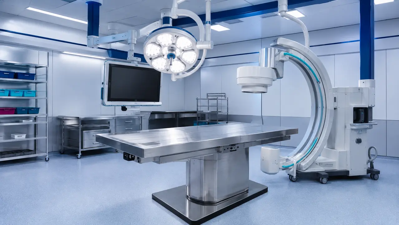
A request for a cadaveric specimen arrives with a clinical objective, but the operational reality of fulfilling that request begins with donor provenance and Ministry of Health supervision. Course directors and medical device engineers often evaluate a facility based on equipment lists, yet the deciding factor for a successful study is the documentation chain supporting the anatomical material.
Our facility operates from Science Park, Rehovot, managing the full operational sequence from donor recruitment and pre-scan imaging to the proper treatment of the remains. We import specimens fresh-frozen after procurement according to the research need, matching donor age, weight, BMI and clinical conditions to the target patient population.
Understanding the distinction between general anatomical separation and procedure-focused validation determines whether a session validates a surgical approach or merely demonstrates it.
Reading time: 9 minutes
Key points
- Human dissection provides tactile and visual accuracy for appreciating natural anatomical variability that synthetic models cannot replicate.
- Cadaver surgical training replicates the operative environment, utilizing surgical instruments, imaging integration, and complete surgical teams.
- Consent-based donation requires strict governance, with chain-of-custody documentation tracking the specimen from procurement through final disposition.
- Defensible donor-based research and development requires protocols, objectives, data capture plans, and strict governance alignment.
Contents
- What is human dissection in medicine?
- Why is human dissection still used in surgical education?
- How is cadaver dissection different from prosection?
- What is cadaver surgical training (CST)?
- How are human body donors governed?
- What ethical standards define a donor-based lab?
- What safety controls are standard in dissection environments?
- How do you select an anatomy training modality?
- How does dissection support medical device R&D?
- What should a course director include in a lab curriculum?
- How do you measure outcomes from cadaver dissection?
- What are common compliance pitfalls in donor programs?
What is human dissection in medicine?
Human dissection is the structured anatomical separation and study of human donor tissues under controlled conditions. The purpose is to understand the spatial relationships between structures, support medical education, and facilitate procedural training. Cadaveric dissection provides a tactile and visual accuracy that synthetic models cannot fully replicate, allowing trainees to appreciate natural anatomical variability.
General anatomy learning and procedure-focused training overlap but serve distinct goals. General education focuses on broad structural recognition, while clinical anatomy training targets specific surgical approaches and instrument pathways. The American Association for Anatomy provides operational definitions and educational context for utilizing cadaveric material, emphasizing that structured engagement with human tissue remains fundamental to medical comprehension.
When a specimen is prepared for a specific otolaryngology or spinal procedure, the dissection plan changes. The approach dictates which structures are preserved and which are reflected. This tailored preparation ensures the specimen yields the required procedural insights without compromising the surrounding anatomy.
Why is human dissection still used in surgical education?
Human dissection remains a high-fidelity method for learning three-dimensional anatomy and rehearsing surgical approaches. In a surgical skills lab, donor-based training allows teams to practice approach selection, instrument handling, and team choreography. Imaging-guided steps can be rehearsed with actual tissue feedback, which is critical for procedures requiring precise spatial awareness.
Peer-reviewed perspectives on the enduring value of donor-based education in modern curricula confirm that translational anatomy research bridges the gap between theoretical knowledge and clinical application. A hybrid anatomy curriculum often combines digital visualization with hands-on donor tissue practice, optimizing the learning sequence for complex procedures.
When a surgical team rehearses a minimally invasive approach, they encounter tissue plane resistance and anatomical variations that virtual reality cannot simulate. The physical constraints of the operative corridor, the behavior of the instruments against actual tissue, and the integration of fluoroscopy create a realistic operative environment. This realism is why donor-based rehearsal remains a standard for high-stakes procedural training.
How is cadaver dissection different from prosection?
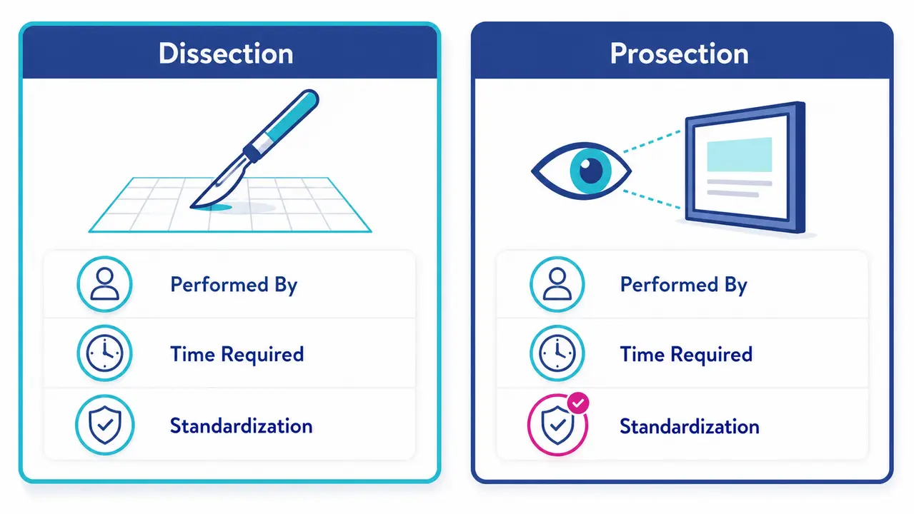
The distinction between prosection vs dissection lies in who performs the tissue separation. Dissection is performed by learners under supervision, requiring them to navigate tissue planes and identify structures sequentially. Prosection is prepared by experienced anatomists for demonstration, allowing learners to observe complex or difficult-to-expose structures without performing the separation themselves.
The choice between the two depends on time-limited curricula, assessment formats, and standardization needs. Prosection offers consistent anatomical exposure across multiple stations, ensuring every trainee sees the same pathology or structural variation. Dissection consumes more time but develops different cognitive and motor skills.
When does prosection outperform dissection for teaching efficiency?
Prosection is superior for standardized demonstrations, limited lab hours, and consistent exam preparation. When the objective is rapid recognition of a complex anatomical region, such as the temporal bone, pre-prepared specimens allow trainees to study the target structures directly without expending hours on removal of overlying tissue.
When does learner-performed dissection add unique value?
Learner-performed dissection develops procedural thinking, step sequencing, and hands-on spatial reasoning. Navigating a tissue plane requires the trainee to adapt to resistance and unexpected anatomical variations. This process builds the cognitive framework necessary for live operative surgery, where tissue behavior is unpredictable.
What is cadaver surgical training (CST)?
Cadaver surgical training (CST) is procedure-focused training using human donors, organized around clinical workflows and endpoints. Unlike general dissection, CST replicates the operative environment, utilizing surgical instruments, imaging integration, and complete surgical teams. The focus is on rehearsing a specific procedure from incision to closure.
Standard CST components include a pre-brief, defined objectives, station design, imaging integration, a structured debrief, and competency-oriented assessment. Scientific literature defines CST as a high-stakes simulation method that validates both individual technical skills and team-based procedural workflows. Imaging-integrated cadaver training allows surgeons to verify screw placement or implant positioning in real-time using C-arm fluoroscopy.
Competency-based surgical education relies on these structured sessions to move beyond simple task repetition. A CST station must replicate the operative field accurately. When a team rehearses a spinal fusion, they require the same instrumentation, imaging support, and sequential workflow they will use clinically. The cadaveric specimen provides the tissue fidelity necessary to validate the approach.
How are human body donors governed?
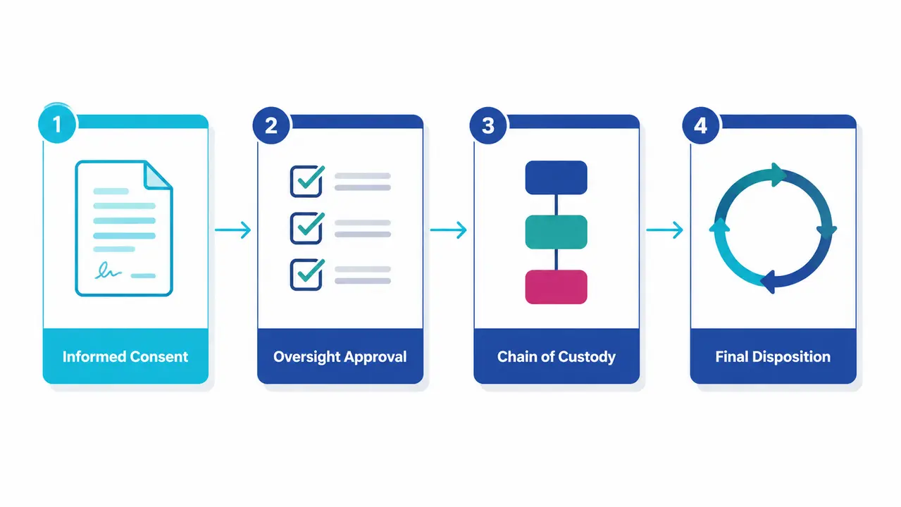
Consent-based donation involves informed permission for educational or research use, supported by institutional governance and documentation. Body donor program governance requires that the scope of use authorized by the donor is strictly followed. A donor consenting to anatomical education may not automatically authorize medical device development research.
Oversight committees and regulatory authorities establish the ethical framework for donor-based education. The Human Tissue Authority provides governance standards and consent codes that define authorization scope, permitted uses, and auditability requirements. Chain-of-custody documentation tracks the specimen from procurement through final disposition, ensuring every handling step is recorded.
Our facility operates under Ministry of Health supervision. Donor ID information, including age, weight, BMI, clinical conditions, and prior surgeries, is documented and available as long as permitted by law and donation conditions. This documentation ensures the donor match is appropriate for the research question and that the ethical boundaries of the donation are respected.
What ethical standards define a donor-based lab?
Ethical practice in a donor-based anatomy lab centers on respect, confidentiality, professionalism, and using donor gifts strictly for approved purposes. Anatomy lab professionalism is not a passive stance but an enforced operational standard. A statement of respect translates into specific behavioral controls and facility protocols.
Medical breakthroughs became possible due to body donors. We honor this by maintaining a sterile, tidy, and respected working environment, with a professional team handling the whole process and safety officers guaranteeing best practice according to guidelines. The International Federation of Associations of Anatomists provides ethical recommendations for good practice around human tissue image acquisition and use.
What should a statement of respect cover operationally?
Operationally, a statement of respect mandates strict access control, a clear photography policy, enforced language standards, meticulous documentation, and defined end-of-use processes. Unauthorized imaging of specimens is prohibited. Access is restricted to authorized personnel directly involved in the approved educational or research activity.
How do course directors reinforce professionalism consistently?
Course directors reinforce professionalism through orientation, faculty modeling, defined incident pathways, and assessment alignment. Professionalism is assessed alongside technical skills. If a trainee breaches conduct standards, the incident pathway requires documentation and remediation.
What safety controls are standard in dissection environments?
Safety in human dissection environments relies on engineering controls, administrative controls, and personal protective equipment aligned to preservation methods and risk assessments. Anatomy lab safety protocols dictate how specimens are stored, handled, and disposed of. Ventilation controls in anatomy labs are critical when working with preservatives or fresh-frozen tissue.
Adherence to established rules for cadaver lab environments is foundational to operational safety. Gross anatomy laboratory standards require that facilities evaluate local extraction at tables, implement air-change strategies, and conduct preservative exposure monitoring. The Occupational Safety and Health Administration provides guidelines for formaldehyde exposure, establishing limits and medical surveillance requirements.
What questions should you ask about ventilation?
Course leadership should verify local extraction at tables, the air-change strategy, monitoring approaches, and maintenance documentation. A facility must provide airflow testing records and chemical monitoring logs to prove the engineering controls function as designed.
What PPE expectations are typical for professional labs?
Standard PPE includes eye protection, gloves, protective garments, and respiratory protection policies based on risk assessment. When a procedure involves aerosolization or specific chemical use, respiratory protection requirements escalate based on the hazard analysis.
How do you select an anatomy training modality?
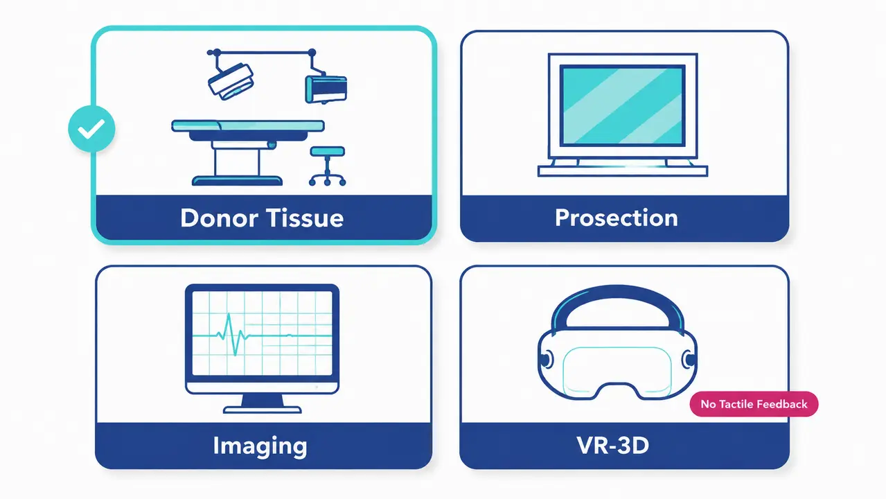
Modality selection depends on learning objectives, required realism, standardization needs, time constraints, and assessment strategy. If the objective is approach rehearsal with imaging, donor-based CST is prioritized. If rapid recognition is the goal, prosection or imaging-based anatomy may suffice. The decision logic hinges on what the training must validate.
A synthetic model cannot reproduce tissue plane variability, so it cannot validate a dissection step. Virtual reality platforms offer procedural sequencing but lack tactile feedback. A hybrid approach often yields the most robust educational outcomes by matching each modality to the appropriate learning phase.
| Modality | Best-fit learning objectives | Strengths for procedural training | Limitations / risks | Standardization and assessment suitability | Typical facility requirements | Governance and documentation considerations | Safety considerations | Best use cases |
|---|---|---|---|---|---|---|---|---|
| Dissection | Spatial reasoning, tissue plane navigation | High tactile fidelity, real anatomical variation | Time-intensive, lower standardization | Process-based assessment | Wet lab, ventilation, PPE | Full donor consent and chain-of-custody | Chemical exposure, sharps management | Resident anatomy education, approach development |
| Prosection | Rapid structure recognition | Consistent exposure of complex structures | No hands-on motor skill development | High standardization for exams | Display stations, limited tools | Donor consent for demonstration | Minimal direct handling risks | Review sessions, exam preparation |
| Imaging-based anatomy | Radiological correlation | Exact anatomical measurement | No tissue handling or instrument feedback | Highly standardized | CT, MRI, ultrasound workstations | Imaging data governance | No biological material risks | Pre-operative planning, radiology training |
| VR-3D platforms | Step sequencing, cognitive rehearsal | Repeatable, unlimited attempts | Zero tactile tissue resistance | Automated data capture | VR headsets, haptic devices | Software licensing | No biological risks | Initial familiarization, workflow training |
| Hybrid | Comprehensive competency | Combines cognitive and tactile fidelity | Complex scheduling and cost | Multi-modal competency metrics | Full wet lab and simulation center | Integrated governance across modalities | Combined engineering and PPE controls | Advanced procedural courses, device validation |
Which objectives map best to each modality?
Anatomy recognition maps efficiently to prosection and imaging. Variability appreciation requires donor-based dissection. Procedural sequencing is initially trained on VR platforms before transitioning to cadaveric tissue for device handling and team-based workflow validation. The objective dictates the fidelity required.
How does dissection support medical device R&D?
Human dissection supports early feasibility, usability evaluation, approach development, and training content creation when realistic anatomy is required. Medical device usability evaluation depends on observing how an instrument interacts with human tissue. Donor-based anatomical models support medical device testing workflows prior to clinical studies.
R&D teams utilize fresh-frozen specimens to test instrument reach, access corridors, imaging visibility, and workflow timing. A device engineered for a minimally invasive cardiac procedure must navigate the anatomical constraints of the thoracic cavity. Testing on a human specimen reveals design constraints that synthetic bench models obscure.
What documentation makes donor-based R&D defensible?
Defensible donor-based R&D requires protocols, objectives, data capture plans, and governance alignment. The research record must demonstrate that the specimen use fell within the consent scope. Study protocols define the endpoints, while chain-of-custody documentation tracks specimen handling. Data capture plans ensure that usability metrics are recorded systematically.
What should a course director include in a lab curriculum?
A strong curriculum defines measurable objectives, faculty-to-learner ratios, station design, time-on-task, prerequisites, and structured debriefs. Procedure lab curriculum design begins with the end goal. If the objective is for a trainee to independently perform a specific orthopaedic approach, the curriculum must sequence the steps logically.
A course director anatomy lab checklist includes pre-reading, anatomy review, step lists, error modes, imaging checkpoints, and post-lab evaluation. These curricula are implemented within specialized medical education laboratories to ensure optimal learning environments. Station design dictates the equipment available, such as C-arm fluoroscopy or laparoscopic towers, and the specimen type required.
Faculty-to-learner ratios are tighter for advanced procedural training. A complex spinal reconstruction requires close supervision to ensure correct technique and patient safety principles. The curriculum must allocate sufficient time-on-task for the trainee to repeat the critical steps until competency is achieved.
Plan your next procedural training session
Tell us what your study needs and we will match the specimen, imaging and staffing to your protocol.
How do you measure outcomes from cadaver dissection?
Outcomes from cadaver dissection or CST must match the intent of the training, focusing on knowledge gains, procedural performance metrics, and calibrated confidence. Measuring satisfaction alone is insufficient for professional training validation. Validated assessment tools provide objective data on skill acquisition.
Practical metrics include checklists, global rating scales, time-to-complete steps, error counts, and retention follow-ups. Scientific literature outlines validated methodologies for evaluating surgical skills, emphasizing that performance metrics must correlate with clinical competency. A procedure-specific checklist verifies that critical steps were completed safely.
Global rating scales evaluate the quality of the motion and tissue handling. Time-to-complete metrics establish efficiency, while error counts flag safety deficiencies. Retention follow-ups, conducted months after the session, determine if the skill was consolidated or if refresher training is required.
What are common compliance pitfalls in donor programs?
Common compliance and operational pitfalls in donor-based dissection programs include unclear consent scope, weak documentation, inconsistent professionalism enforcement, inadequate safety controls, and poor separation of education versus research activities. Donor-based anatomy education requires strict boundaries between learning activities and product development research.
Prevention methods include governance reviews, standard operating procedures, audit trails, incident reporting, and clear boundaries for data capture. When a session shifts from teaching a surgical approach to collecting data for a device patent, the consent scope and governance approvals must support that transition.
| Process Stage | Operational Node | Required Action |
|---|---|---|
| Entry point | Proposed activity | Define educational or research objective |
| Decision node | Consent scope alignment | Verify donor authorization permits the activity |
| Decision node | Oversight approval | Secure ethics committee or institutional approval |
| Decision node | Documentation required | Establish chain-of-custody and protocols |
| Decision node | Data capture rules | Define imaging and metric recording boundaries |
| Operational control | Access control | Restrict entry to authorized personnel |
| Operational control | Professionalism | Enforce statement of respect and conduct standards |
| Operational control | Safety controls | Verify ventilation and PPE compliance |
| Output | Training outcomes | Collect assessment metrics |
| Output | Research records | Archive data capture and usability results |
| Output | Audit trail | Compile all governance and operational logs |
| Close-out | End-of-activity documentation | Finalize records and confirm proper treatment of remains |
| Close-out | Debrief | Review errors and outcomes with the team |
| Close-out | Continuous improvement loop | Update protocols based on session findings |
Frequently asked questions
When is prosection the better choice than dissection?
When time is constrained and standardisation matters more than discovery. Prosection is prepared in advance by experienced anatomists, so every trainee at every station sees the same structure or variation. Dissection consumes considerably more time but develops the sequential tissue-handling skill that prosection cannot teach. Time-limited curricula and assessment formats usually decide it.
What does consent scope actually restrict?
It restricts the purposes a donor may be used for, not merely the fact of use. A donor who consented to anatomical education may not have authorised medical device research. Body donor programme governance requires that the authorised scope is followed strictly, and chain-of-custody documentation has to demonstrate that the actual use stayed inside it.
What changes when a teaching session starts capturing device data?
The governance basis changes. When a session shifts from teaching a surgical approach to collecting data supporting a device claim, the consent scope and the governance approvals must already support that transition. Deciding this mid-session is one of the most common compliance failures, so the boundary between education and research activity is set before the lab begins.
Which metrics actually validate procedural training?
Satisfaction scores alone are insufficient. Practical measures include structured checklists, global rating scales, time-to-complete for defined steps, error counts and retention follow-up. The measures have to match the intent of the training, so a session aimed at approach selection is assessed differently from one aimed at instrument handling.
What ventilation and exposure controls does a dissection environment need?
Facilities evaluate local extraction at the tables themselves, implement an air-change strategy for the room, and monitor preservative exposure. These engineering controls sit alongside administrative controls and personal protective equipment, and the combination is matched to the preservation method in use and to the risk assessment for that method.
Selecting the right anatomical modality, enforcing operational professionalism, and maintaining strict governance boundaries determine whether a cadaveric session validates a surgical approach or merely demonstrates it. Our facility provides the fresh-frozen specimens, imaging integration, and Ministry of Health supervision required to ensure your study or course meets its clinical objective.
About Biotech Anatomy

Biotech Anatomy LTD has provided practical anatomy and surgical education from Science Park, Rehovot since 2017. We are the only private Israeli company dedicated to employing human cadaveric material for research, and we operate fully equipped surgery rooms and classrooms with advanced monitoring, imaging and surgical equipment. We support medical teams, academic researchers and medical device developers through the whole study: donor recruitment and matching to the research need, pre-scan imaging, import and preparation of the specimen, trained staff throughout the session, and proper treatment of the remains afterwards, all under Ministry of Health supervision. Specimens are imported fresh-frozen after procurement, and donor information and diagnostic data are made available as far as law and the terms of donation permit. Our facility exists to honour what body donors intended their donation to achieve.