Cadaver Dissection in Medical School
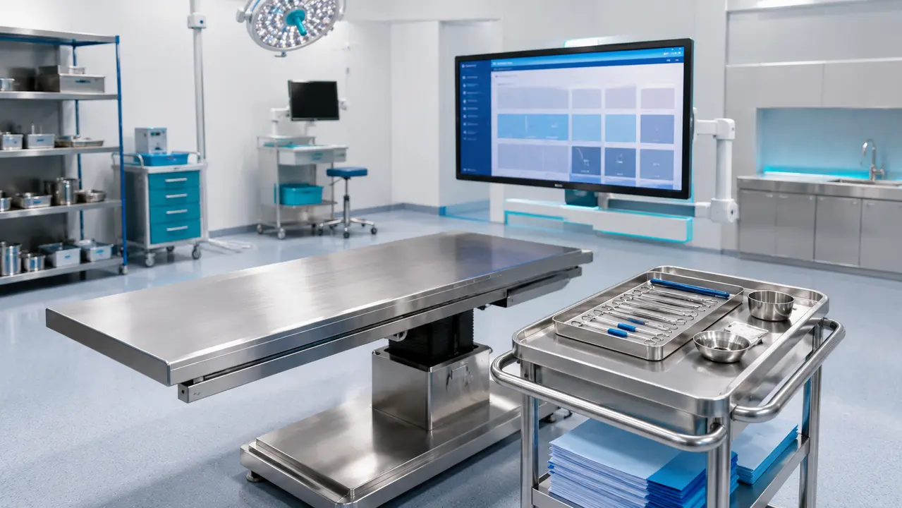
Faculty supervising a modern anatomy block must reconcile three competing pressures: limited curriculum hours, the demand for high-fidelity spatial reasoning, and strict institutional governance. Cadaver dissection in medical school remains the primary mechanism for teaching these three-dimensional relationships, anatomic variation, and professional practice behaviors. This is structured, faculty-supervised anatomical study using donated human bodies. Modern programs integrate dissection within specialized medical education laboratories rather than treating it as an isolated exercise.
Dissection is one element within a broader anatomy learning system that includes imaging, models, and simulation. Educators and procedural specialties value the competency outcomes it produces: spatial reasoning, tissue-plane logic, team communication, and disciplined lab conduct. A donor-based anatomy education framework positions the specimen as the central reference point for these competencies. The cadaveric dissection curriculum requires learners to navigate real tissue variability, which synthetic models cannot fully replicate. By progressing through regional anatomy blocks, learners build a variation-aware mental map that directly supports later clinical decision-making.
Reading time: 9 minutes
Key points
- Cadaver dissection provides tactile feedback and real anatomical variability that synthetic models and virtual reality cannot replicate.
- Strict lab orientations, timeboxing, and safety protocols are required to ensure professional conduct and sharps discipline.
- Ethical stewardship governs every aspect of the process, ensuring donor consent and privacy boundaries are respected.
- Specimen selection is a deliberate design decision based on the specific clinical or surgical learning objectives.
Contents
- How dissection differs from prosection teaching
- Why imaging and simulation cannot replace dissection
- Designing a first dissection orientation
- Standard PPE and safety practices
- Managing ventilation and chemical exposure
- Professionalism standards in the dissection room
- Ethical stewardship and respect for body donors
- Structuring lab time for dissection sessions
- Typical learning objectives for dissection sessions
- Assessing competence after cadaver dissection
- When cadaveric training is preferred beyond medical school
- Comparison of instructional and training models
- Specimen selection as a design decision
How dissection differs from prosection teaching
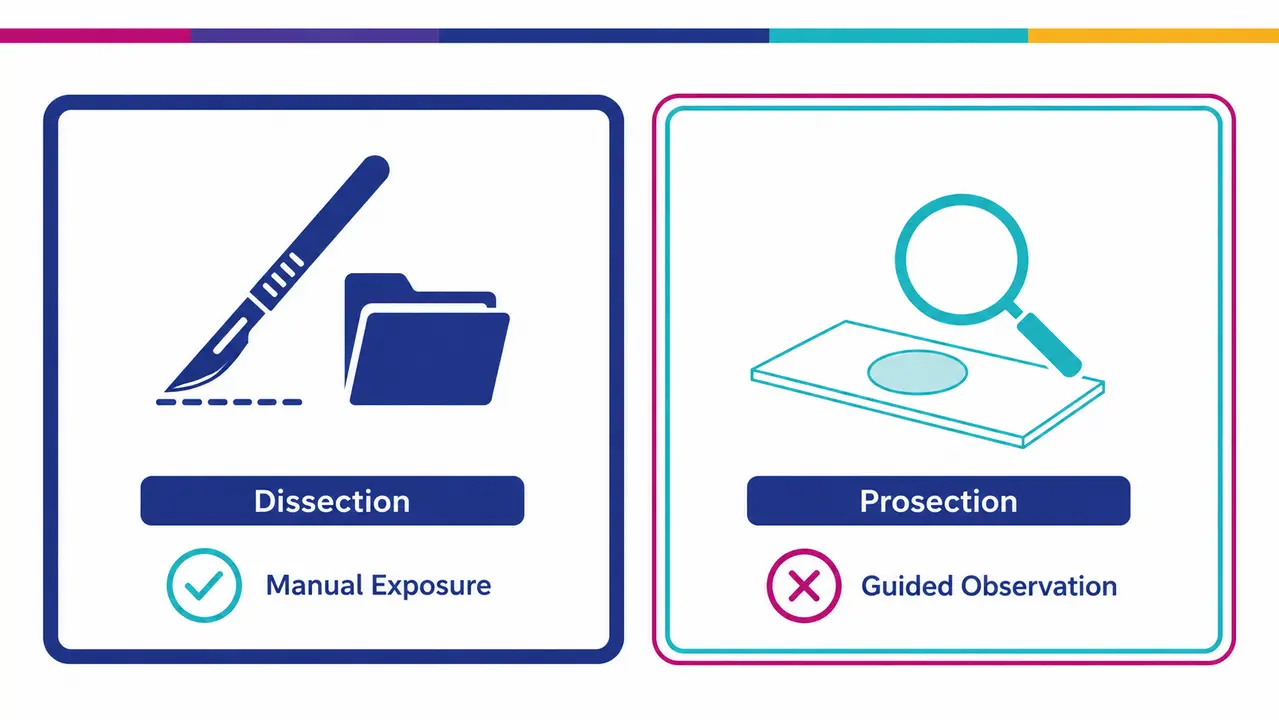
Course directors deciding between formats must weigh preparation time against observation time. In dissection, learners perform stepwise exposure under supervision. In prosection, learners study professionally prepared specimens. The choice between prosection vs dissection alters the instructional design, staffing needs, and how the session supports an anatomy practical exam.
Prosection as an instructional choice
Prosection involves faculty preparing specimens in advance to display specific structures. It is often chosen when time efficiency is critical and the focus is purely on identification and clinical correlation rather than manual exposure skills. Under faculty supervision gross anatomy teaching shifts from manual discovery to guided observation. This format allows high-volume review of complex regions in a compressed timeframe.
Dissection and unique educational value
Dissection adds unique value when tactile feedback, tissue plane navigation, and discovery of anatomic variability are the primary learning objectives. Learners trace neurovascular structures through fascia, encountering real anatomical deviations that prepared specimens often obscure. Adherence to a strict anatomy lab SOP ensures this manual exploration remains systematic. The process forces learners to make real-time decisions about tissue removal based on structural relationships.
Why imaging and simulation cannot replace dissection
Imaging, virtual reality, and simulation provide excellent supplementary training but lack the physical constraints of real tissue. A virtual reality model cannot teach a learner how much force is required to retract a nerve without causing traction injury. Dissection remains valuable because it teaches realistic spatial relationships and anatomic variability in a controlled, respectful environment. This variation-aware decision-making is crucial for surgeons and interventionalists who will eventually navigate unexpected anatomy in a living patient.
Programs integrate dissection with imaging review, specifically CT and MRI correlation, to bridge the gap between gross anatomy and radiological interpretation. Structured reflection on donor stewardship also contributes to professional identity formation anatomy. Students learn to manage the emotional and ethical weight of working with human remains, which builds the discipline required in clinical practice. This clinical correlation anatomy education ensures that learners understand the direct link between a dissected structure and a patient presentation. National curriculum reports consistently show that medical school curricula retain dissection precisely because digital tools cannot replicate this combination of tactile reality and professional accountability.
Designing a first dissection orientation
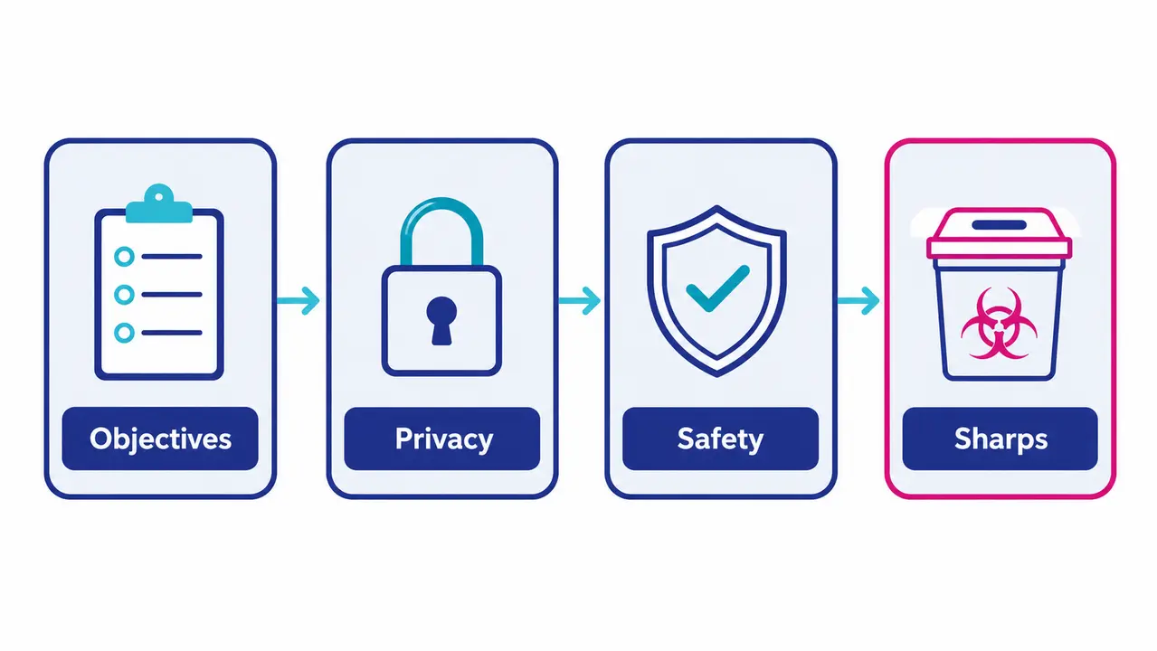
A strong cadaver lab orientation aligns learning objectives, safety controls, and professionalism expectations before hands-on work begins. The operational checklist must cover lab flow, assign specific roles to faculty, the lab manager, and the safety officer, and define permitted materials. Orientation also establishes photography rules and escalation pathways for incident reporting. Clear boundaries at this stage prevent disruptions later.
Pre-brief objectives and timeboxing
The pre-briefing process requires course directors to clearly state the learning objectives for the session. Directors must assign team roles, designating who will dissect, who will assist, and who will read from the anatomical guide. Establishing strict time boundaries for each dissection step keeps the cohort aligned. Timeboxing prevents groups from spending an entire session on a single region while neglecting subsequent structures.
Professional conduct and privacy boundaries
Expected professional conduct in the lab mirrors the standards of a clinical environment. Instructors must emphasize the use of respectful donor terminology rather than casual references. Strict prohibitions on unauthorized photography form the core of the specimen privacy and photography policy. Learners must also understand clear boundaries on what can be documented in personal notes. Maintaining anatomy lab professionalism ensures the dignity of the donation is preserved throughout the educational process.
Safety brief and sharps discipline
Before any instruments are distributed, instructors must verify personal protective equipment compliance. The safety brief reinforces sharps safety anatomy lab protocols, specifically addressing the handling and disposal of scalpels and needles. Instructors must demonstrate the correct technique for passing a sharp instrument and establish the location of designated sharps containers. Enforcing these rules before the first incision prevents preventable exposure incidents.
Standard PPE and safety practices
Safety protocols in a cadaveric facility follow local SOPs to mitigate biological and chemical risks. Standard PPE for anatomy lab usage dictates that all personnel wear protective gloves, eye protection, and fluid-resistant gowns or aprons. Double-gloving is often required when learners are performing active dissection, allowing the outer glove to be changed if it tears or becomes heavily contaminated. The selection of equipment must be fit for purpose rather than tied to specific brand recommendations. Gross lab safety procedures emphasize training and proper doffing techniques to prevent contamination of clean areas.
Personnel must remove PPE in a specific sequence to avoid transferring preservatives or biological materials to the skin or clothing. The facility provides the necessary safety infrastructure, including eye-wash stations and emergency showers. Trained staff monitor compliance throughout the session. A professional team handles the whole process, with environmental and safety officers guaranteeing best practice according to guidelines. This operational rigor ensures that the working environment is kept sterile, tidy and respected.
Managing ventilation and chemical exposure
Labs manage exposure risk through engineering controls and defined workflows. Course directors should ask facilities teams about ventilation targets, air monitoring schedules, and room hygiene protocols. Engineering controls ventilation anatomy lab standards dictate specific air exchange rates to remove chemical vapors. Chemical exposure control anatomy lab procedures ensure that vapor concentrations remain below established occupational limits.
Engineering controls and room design
Room design for anatomical facilities requires adequate air exchange rates to maintain safe breathing zones. Downdraft tables are utilized where applicable to draw vapors away from the user before they enter the room air. Proper waste disposal infrastructure must exist for both chemical and biological materials to prevent accumulation at workstations.
SOPs for chemical handling and exposure response
Standard operating procedures govern the handling of all preservatives and chemical agents used in the facility. General chemical safety protocols are maintained without citing specific OSHA exposure limit numbers, focusing instead on the existence of continuous monitoring and spill response protocols. Facilities must comply with occupational exposure to formaldehyde standards to protect personnel.
Access control and chain of custody basics
High-level chain of custody anatomical specimens protocols track the movement of every donor from intake to final disposition. Access control anatomy lab rules ensure that only authorized personnel are present in the facility. Logging access creates an auditable record of who interacted with the specimen and when.
Professionalism standards in the dissection room
Professionalism in the dissection room mirrors clinical professionalism. The environment operates like an operating room where disciplined team behaviors are mandatory. No identifying details regarding the donor may be discussed or documented outside the authorized educational context. Controlled photography policies prohibit any imaging unless it is explicitly authorized for educational archives. Appropriate language is enforced strictly. Faculty model respectful communication, correcting any casual or dismissive references to the human specimen. The cadaveric dissection curriculum evaluates these behaviors alongside anatomical knowledge. A learner who identifies a structure perfectly but handles the tissue carelessly fails the professional standard. This expectation extends to workspace organization. Teams must maintain a clean station, dispose of tissue remnants in designated containers, and treat the remains with consistent dignity. The transition from student to physician requires this early internalization of clinical discipline.
Ethical stewardship and respect for body donors
Ethical stewardship of donors is demonstrated through consent-aware use, respectful handling, privacy protection, and transparent governance. A human body donor program operates on the premise that individuals have consented to their use for specific educational or research purposes. The facility enforces a clear statement of respect (donor respect statement) that governs all interactions with the specimens. Medical breakthroughs and advancements in medicine along history became possible due to body donors. This ethical position is grounded in operations: the working environment is kept sterile, a professional team handles the whole process, and environmental and safety officers guarantee best practice according to guidelines. Donor acknowledgement ceremonies, faculty modeling respectful language, restricted access, and clear boundaries on secondary use reinforce this educational impact of dissection. Biotech Anatomy operates under Ministry of Health supervision to ensure these standards are met without compromise.
Consent-aware scope of use and governance
The scope of use, whether for education, research, or training, must align strictly with the donor documented consent. Institutional governance boards oversee this alignment to prevent unauthorized utilization. A specimen consented for gross anatomy education cannot be repurposed for device development without explicit permission.
Privacy imagery and publication boundaries
Any imagery used for educational publication must be strictly de-identified. Images must comply with institutional and national tissue authority guidelines regarding donor privacy. Identifying features must be obscured or excluded from the frame entirely to maintain anonymity.
Acknowledgement practices that reinforce respect
Acknowledgement practices avoid being performative. Faculty lead silent reflection or state a brief acknowledgement of respect at the start of a block. Institutional gratitude is demonstrated through the careful, purposeful use of the tissue rather than through excessive ceremony.
Structuring lab time for dissection sessions
The duration of cadaver dissection in medical school varies widely across institutions. Most programs structure dissection into repeated, timeboxed sessions aligned to regional anatomy blocks and assessment milestones. Common scheduling patterns include weekly labs, alternating groups, and hybrid dissection and prosection models. Some institutions run full-day immersive sessions, while others prefer shorter, focused two-hour blocks. The planning logic depends on faculty availability, lab capacity, and the overall curriculum structure.
Directors must sequence the progression logically. A typical sequence begins with the back, progresses through the thorax and abdomen, and concludes with the head and neck or extremities. This progression mirrors the regional anatomy blocks taught in didactic lectures. Assessment milestones dictate the pacing. A practical exam scheduled at the end of a block requires that all relevant structures be exposed and reviewed beforehand. Programs often use a hybrid approach where prosection is used for regions where dissection is overly complex or time-consuming, while full dissection is reserved for high-yield areas. The facility accommodates these varying schedules by providing fully equipped surgery rooms and classrooms. Our clients requiring torso, cephalous, pelvic or extremities specimens are served with the necessary technical support to execute their specific curriculum design.
Typical learning objectives for dissection sessions
Typical objectives for a practical session use measurable verbs: identify, differentiate, trace, and correlate. Learners must identify major neurovascular structures, differentiate between adjacent muscle groups, trace nerve plexuses from origin to target, and correlate anatomical findings with clinical conditions. These objectives directly prepare learners for the anatomy practical exam. Advanced objectives for professionals include approach planning and risk structure awareness. A surgical resident must demonstrate the ability to plan a safe surgical approach to a specific structure while avoiding critical adjacent tissues.
Identification and relationship mapping
Learners must demonstrate the ability to map three-dimensional spatial relationships between adjacent structures. This requires understanding how a nerve passes deep to a specific artery and superficial to a named muscle. Three-dimensional understanding prevents accidental injury during clinical procedures.
Variation awareness and clinical correlation
Learners must recognize normal anatomic variations and correlate them with clinical findings or imaging. A muscle variant might explain an unusual presentation of nerve compression. Recognizing these variations builds the pattern recognition required for safe surgical practice.
Team-based workflow and communication objectives
Learners are evaluated on their ability to communicate effectively within a team. Tasks are delegated clearly, and team members maintain a sterile, organized workspace. Effective communication in the lab translates directly to safer operating room dynamics.
Assessing competence after cadaver dissection
Assessment of anatomical competence commonly combines timed identification, applied clinical questions, and structured evaluation of professionalism. Timed identification, often called a spotter exam anatomy format, requires learners to quickly name tagged structures on a specimen. OSPE anatomy stations test applied reasoning by asking learners to identify a structure and explain its clinical significance or trace its proximal origin. These assessments must align directly with the dissection learning objectives.
Educators design rubrics to capture multiple dimensions of competence. Accuracy in identification forms the baseline. Reasoning evaluates whether the learner understands the functional and clinical relevance of the structure. Safe technique assesses how the learner handles tissue and instruments. Respect compliance monitors adherence to lab rules and professional conduct. A learner who identifies a structure but damages surrounding tissue to expose it loses points on safe technique. Remediation pathways address specific deficits. If a cohort struggles with spatial relationships, directors schedule additional imaging correlation sessions. Feedback loops close the gap between assessment and instruction. Faculty review the practical exam results to identify common misconceptions and adjust the next dissection block accordingly. This continuous improvement ensures the curriculum remains responsive to learner needs.
When cadaveric training is preferred beyond medical school
Cadaveric training is preferred beyond medical school when realistic anatomy, variation, and full procedural workflows are essential for skill transfer, protocol rehearsal, or device evaluation before clinical use. Surgeons, CME leads, and medtech teams require human tissue to validate procedural steps that synthetic models cannot support. Cadaveric surgical training allows surgeons to rehearse new techniques, evaluate instrumentation ergonomics, and practice imaging-guided workflows. This cadaveric training often complements surgical simulation training by providing the final layer of biological fidelity. Translational research in a translational research anatomy lab supports medical device testing by allowing engineers to evaluate a device pathway in human anatomy. A CME cadaver lab provides the environment for continuing education, while a medical device R&D cadaver lab facilitates pre-clinical evaluation.
CME and advanced procedural rehearsal use cases
CME courses utilize fresh-frozen specimens for realistic tissue feedback during procedural rehearsal. The focus is on technique refinement rather than basic identification. Procedural skills lab sessions rely on fresh-frozen vs embalmed donors to provide realistic tissue pliability for tasks like vessel anastomosis or joint replacement. Cadaveric simulation training bridges the gap between dry lab practice and live surgery.
Medical device R&D and translational evaluation use cases
Device teams use cadaveric models to evaluate instrumentation ergonomics and device pathways in human anatomy. Engineers test whether a new instrument can reach a target structure without impinging on surrounding tissue. These sessions do not make regulatory or performance claims but provide critical pre-clinical design feedback.
Governance and documentation for advanced sessions
Advanced R&D and CME sessions require stringent documentation of specimen provenance, consent scope, and access control. These standards mirror academic lab requirements. Every device tested and every procedure rehearsed must be logged against the specific donor consent parameters to maintain ethical compliance.
Comparison of instructional and training models
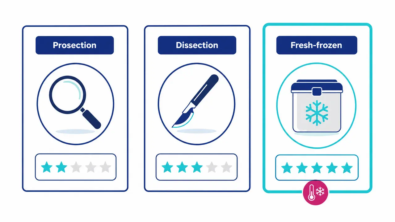
| Model | Primary Use Case | Tissue Fidelity | Key Constraint |
|---|---|---|---|
| Prosection | High-volume identification and review | Fixed, professionally prepared | No manual exposure skill development |
| Dissection | Spatial reasoning and tissue plane logic | Fixed or fresh-frozen | Requires significant faculty supervision |
| Fresh-frozen Cadaveric Lab | Surgical rehearsal and device R&D | High, mimics living tissue | Strict cold-chain management required |
Specimen selection as a design decision
Specimen selection is a design decision driven by the clinical question. The clinical question determines the specimen type, condition, and imaging requirements. A course teaching minimally invasive spine techniques requires a different specimen than a course teaching superficial plastic reconstruction. Biotech Anatomy both helps in donor recruitment and matching the most appropriate donor for the research needs. Donor ID information, including age, weight, BMI, and clinical conditions, is available as long as permitted by law and donation conditions. Diagnostic tools, including X-ray, MRI, genetic information for mutations, hereditary information, and former surgeries, are also documented.
Pre-scan imaging, specifically CT or MRI, is ordered before the specimen is scheduled for the session. This allows course directors to verify that the anatomy matches the learning objective. A specimen with severe degenerative joint disease might be ideal for an orthopaedic arthroplasty course but useless for a standard anatomy block teaching normal joint architecture. Organs are imported fresh-frozen after procurement according to research needs. The facility handles the full chain: importing into the facility, preparing the venue for the research, and helping with trained staff all along the study. The remains are treated properly afterwards, all under Ministry of Health supervision. Operating since 2017, Biotech Anatomy is the only private Israeli company dedicated to employing human cadaveric material for research. This integrated chain prevents the gaps in provenance and preparation that compromise study validity.
Tell us what your study needs
Let our laboratory match your curriculum or research objectives with the right specimens and facilities.
Frequently asked questions
What is the educational rationale for cadaver dissection in a modern medical curriculum?
The rationale is based on providing authentic, three-dimensional anatomical variation and tactile feedback that digital models cannot fully replicate. Dissection forces learners to navigate real tissue planes and encounter unexpected anatomical deviations. This process builds the spatial reasoning and variation-aware decision-making required for safe clinical practice.
How do I choose between dissection, prosection, or a hybrid anatomy lab model?
Course directors should base this decision on available faculty staffing, curriculum time constraints, and specific learning objectives. If the objective is pure identification within a limited timeframe, prosection is effective. If manual skill and tissue plane navigation are the goals, dissection is necessary. A hybrid model uses prosection for complex regions to save time.
How do we align anatomy practical assessments with dissection learning objectives?
Assessments must mirror the stated objectives. Use spotter exams for pure identification tasks. Use OSPE stations for applied clinical reasoning and tracing exercises. Implement continuous faculty observation to evaluate safe technique, respect compliance, and team communication during the dissection process itself.
What are common preventable errors in first dissection sessions?
Common errors include inadequate pre-briefing, poor timeboxing, and a lack of clear team roles. Learners often spend too much time on superficial exposure and run out of time for deep structures. These failures are preventable through rigorous orientation, strict time boundaries for each step, and assigning specific dissection roles to every team member.
Cadaver dissection in medical school provides an irreplaceable foundation in spatial reasoning, tissue handling, and professional conduct. Designing a successful program requires strict alignment between learning objectives, safety protocols, and ethical stewardship. Contact our laboratory to discuss how our fully equipped surgical facility and trained staff can support your anatomy curriculum.
About Biotech Anatomy

Biotech Anatomy LTD has provided practical anatomy and surgical education from Science Park, Rehovot since 2017. We are the only private Israeli company dedicated to employing human cadaveric material for research, and we operate fully equipped surgery rooms and classrooms with advanced monitoring, imaging and surgical equipment. We support medical teams, academic researchers and medical device developers through the whole study: donor recruitment and matching to the research need, pre-scan imaging, import and preparation of the specimen, trained staff throughout the session, and proper treatment of the remains afterwards, all under Ministry of Health supervision. Specimens are imported fresh-frozen after procurement, and donor information and diagnostic data are made available as far as law and the terms of donation permit. Our facility exists to honour what body donors intended their donation to achieve.