Integrating cadavers for medical students into anatomy curriculum
A donated human body remains the only model that reproduces the tissue plane variability, structural heterogeneity and spatial complexity of a living patient. Digital platforms can map a cross-section perfectly. They cannot reproduce the mechanical resistance of a fascial layer or the anatomical deviations that alter a surgical approach. Course directors and device engineers must decide what fidelity their specific objective requires before booking a facility.
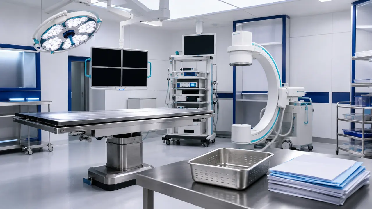
Our facility operates from Science Park in Rehovot. We handle donor recruitment, pre-scan imaging, venue preparation and session staffing under Ministry of Health supervision. The specimens are imported fresh-frozen after procurement, matched to the specific clinical question a study or course is designed to answer.
This article breaks down the operational decisions that govern cadaver-based education and pre-clinical evaluation. It covers specimen selection, imaging correlation, safety governance and the documentation that accompanies a study.
Reading time: 8 minutes
Key points
- Human specimens provide the anatomical variation and tactile feedback that digital platforms and synthetic models cannot reproduce.
- Preservation methods must match the learning objective, with fresh-frozen tissue reserved for procedural rehearsal and device evaluation.
- Consent verification and Ministry of Health supervision govern every stage of donor matching, import and final disposition.
- Blended curricula that pair simulation with cadaveric rehearsal yield the strongest educational and pre-clinical outcomes.
Contents
- What are cadavers used for in medical education?
- Why anatomy curricula retain cadaver-based learning
- How medical schools source human donors
- Preservation methods and specimen selection
- What competencies do cadavers realistically support
- Integrating cadaver teaching into modern curricula
- Cadaver labs versus simulation alone
- Safety and compliance in the cadaver lab
- Ensuring respectful practice and professional conduct
- Donor information and documentation for educational use
- Choosing whole-body donors versus regional donors
- Assessing learning outcomes from cadaver-based anatomy
- Supporting medical device R&D and pre-clinical evaluation
What are cadavers used for in medical education?
Donated human bodies are used to teach three-dimensional anatomy, anatomical variation and clinically oriented procedural skills in a way that aligns closely with real operative and imaging contexts. The objective is not simple memorization. The objective is spatial reasoning, correlation with radiology and supervised skills rehearsal for defined competencies.
Many programs frame the donor as a first patient to reinforce professionalism and respect. This framing is supported by educational research on how cadaver-based anatomy experiences support compassion and professionalism in early training, as detailed by the Association of American Medical Colleges. The donor is treated as a patient, and the learner is held to clinical standards of conduct from the first session.
Our facility applies this framing operationally. A professional team handles the whole process, maintaining a sterile, tidy and respected environment. Safety officers guarantee best practice according to guidelines. The ethical position is stated plainly, then grounded in the physical mechanisms that make it real.
For educators, this means cadavers for medical students are not simply biological teaching aids. They are the medium through which spatial reasoning and clinical professionalism are first tested.
Why anatomy curricula retain cadaver-based learning
Cadaver-based learning uniquely supports tactile-spatial understanding and the appreciation of anatomic variability. Digital tools accelerate repetition and cross-sectional visualization. Human specimens serve as the ground truth for anatomical variation. Digital platforms serve best for pre-lab preparation and post-lab consolidation.
The best educational outcomes typically come from blended design rather than a full replacement of traditional labs. A synthetic model cannot reproduce tissue plane variability, so it cannot validate a specific procedural step. A virtual reality module cannot simulate the mechanical feedback of a real anatomical structure.
Human cadavers in medical school curricula provide the variation that standardized digital models deliberately smooth out. A student who encounters a radicular artery anomaly or a deviated musculotendinous junction during a dissection block learns to adapt to the unexpected. This exposure builds the cognitive flexibility required in an operating room.
An anatomy curriculum that drops the cadaveric component entirely loses this variation exposure. The lab remains the environment where theoretical mapping meets biological reality.
How medical schools source human donors
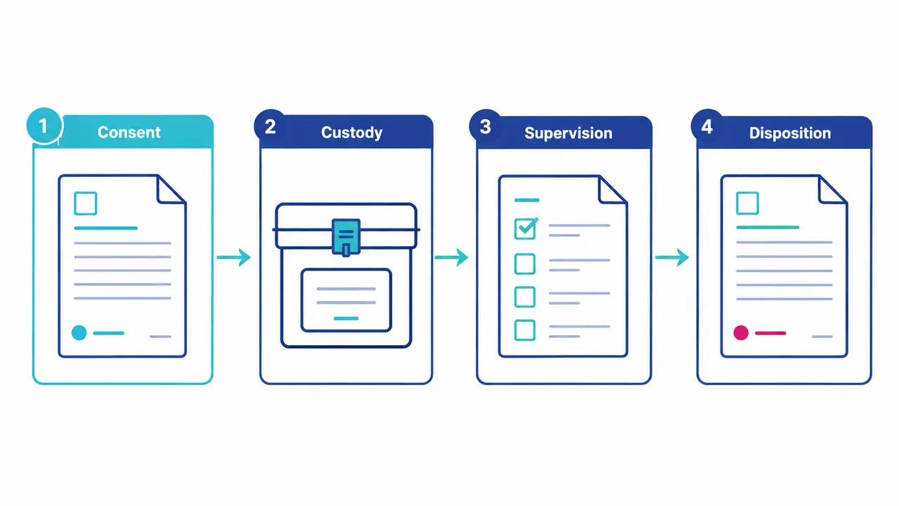
Most institutions rely on regulated body donation programs with documented consent and a controlled chain-of-custody from acceptance through final disposition. Program-based sourcing requires institutional oversight and governance. Educators must verify consent scope, permitted uses and documentation practices before a specimen enters a teaching environment.
Our facility participates in this chain by matching the most appropriate donor for the research needs, importing the specimen under regulatory supervision and treating the remains properly afterward. We operate under Ministry of Health supervision across the entire lifecycle. For a broader look at institutional sourcing mechanisms, course directors can review Where Do Medical Schools Get Cadavers?
Body donation best practices emphasize consent, dignity, governance and legal compliance. The American Association for Anatomy provides consensus recommendations for operating these programs. These standards ensure that a medical school cadaver is procured ethically and utilized within the agreed boundaries.
Consent verification is a hard stop. If a donation agreement limits use to educational purposes, that specimen cannot be diverted to device validation without explicit authorization.
Preservation methods and specimen selection
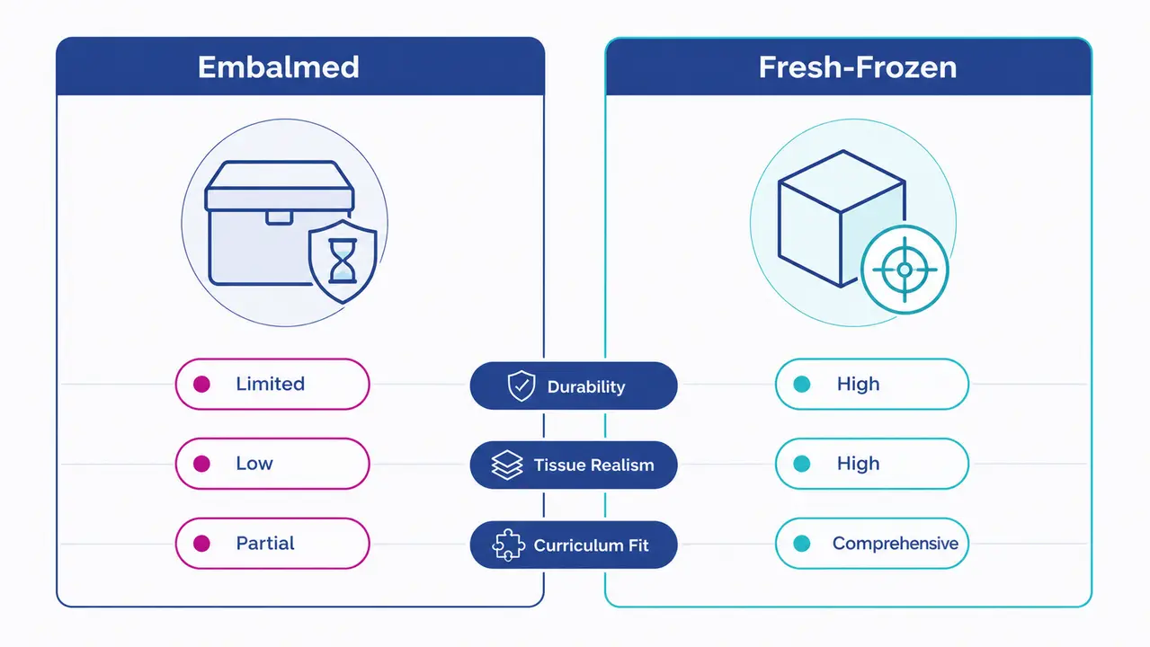
The preservation method affects tissue handling characteristics, flexibility and suitability for specific educational goals. Modality selection should be driven by learning outcomes and safety requirements. Embalmed donors often support longer-term anatomy teaching. Fresh-frozen or lightly preserved models better support procedural rehearsals and device evaluation where tissue realism is critical.
| Modality | Best-fit learning objectives | Typical curriculum placement | Strengths for imaging correlation | Operational considerations | Safety considerations | Documentation needs | Governance checks |
|---|---|---|---|---|---|---|---|
| Embalmed | Structural identification and dissection skills | Pre-clinical | Supports basic CT and MRI correlation | Longer storage duration, flexible scheduling | Chemical exposure controls, formaldehyde monitoring | Donor ID and consent scope | Permitted activities verified |
| Lightly preserved | Procedural introduction and tissue plane identification | Transition | Good for fluoroscopic guidance | Intermediate storage, requires scheduling coordination | PPE emphasis, ventilation engineering | Relevant medical history | Consent for procedural use |
| Fresh-frozen | Procedural rehearsal and device evaluation | CME | High fidelity for imaging and simulated-use | Strict scheduling, specialized storage, intensive staffing | Restricted access, biological hazard protocols | Minimum necessary donor info | Strict scope-of-use alignment |
The table above outlines cadaver modalities for medical education and skills training, selection by learning objective. Selecting the wrong modality invalidates the session. A heavily embalmed specimen will not yield realistic tissue feedback for a laparoscopic tower exercise.
Which modality fits a first-year gross anatomy block
Educators should prioritize durability, scheduling flexibility and consistent access for repeated sessions. A first-year gross anatomy block requires a specimen that can withstand multiple exposures over weeks. Embalmed donors are the standard here because they maintain structural integrity for longitudinal study. A cadaver lab medical school environment must support this repeated access without degrading the educational value of the specimen.
Which modality fits procedure-focused labs and CME courses
Planners should prioritize procedural realism, imaging compatibility and a defined scope-of-use aligned to the donor consent. Our facility provides fresh-frozen specimens for these sessions. Cadavers for medical students in CME courses require the tissue to respond realistically to surgical instruments, C-arm fluoroscopy and laparoscopic equipment.
What competencies do cadavers realistically support
Cadaver-based labs support anatomy identification, variation recognition, imaging correlation and selected foundational procedural skills when objectives, supervision and assessment are explicit. They do not replace supervised patient care. They build the foundational competence required before a learner enters a clinical environment.
These competencies map to specific curriculum phases. Pre-clinical anatomy focuses on structural identification. Transition-to-clerkship introduces procedural skills. Specialty electives use cadavers for focused rehearsals in fields like orthopaedic or otolaryngology. These medical education laboratories provide the supervised skills environments where these competencies are tested.
Our facility supports these phases by providing fully equipped surgery rooms with advanced monitoring and imaging equipment. The facility provides the physical infrastructure. The course director provides the curriculum and the assessment framework.
Integrating cadaver teaching into modern curricula
High-performing curricula integrate pre-reading and imaging, structured lab objectives and post-lab clinical correlation. They do not treat the lab as a standalone experience. A standard workflow begins with pre-lab imaging orientation, moves to in-lab faculty-guided objectives, follows with post-lab case discussion and ends with assessment tied to outcomes.
The following diagram description outlines the cadaver-based anatomy curriculum integration workflow from an educator view. Inputs include learning objectives, competency level, modality selection and governance checks. Pre-lab involves imaging orientation, anatomy learning targets and a safety briefing. In-lab involves structured stations, faculty prompts and documentation of completion. Post-lab involves case-based correlation, imaging review and assessment. Outputs include competency evidence, course QA metrics and a continuous improvement loop.
An anatomy curriculum that fails to connect the lab to clinical practice wastes the resource. The lab must be positioned as a bridge, not an island.
Aligning cadaver sessions with radiology and cross-sectional anatomy
Educators should use standardized imaging sets for correlation and structured prompts for interpretation during the pre-lab and post-lab phases. Cadavers for medical students yield the highest return when the physical specimen is paired with the exact CT or MRI data a clinician would use to plan an approach.
Cadaver labs versus simulation alone
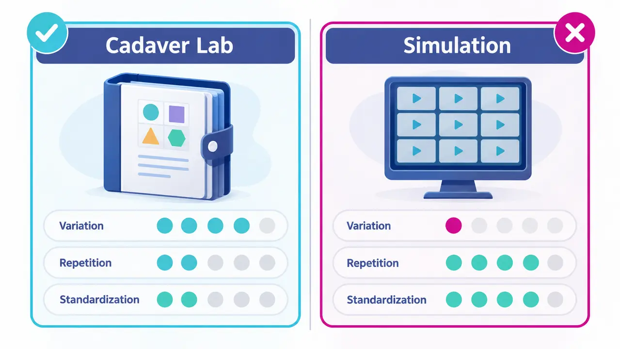
Cadaver labs and simulation address different validity needs. Cadavers strengthen anatomical realism and variation exposure. Simulation improves repetition, standardization and scalable assessment. A synthetic model cannot reproduce tissue plane variability, so it cannot validate a specific procedural step. A cadaver cannot be reset, so it limits repetition.
Educators must choose based on fidelity requirements, risk profile and assessment goals. A cadaver lab medical school session is necessary when anatomical variation is the core learning objective. An anatomy curriculum focused on basic hand-eye coordination might rely more heavily on simulation.
We recommend blended pathways for complex procedures and team training. Combining surgical simulation training with cadaveric rehearsal allows learners to standardize their mechanics before testing their anatomical adaptability.
Safety and compliance in the cadaver lab
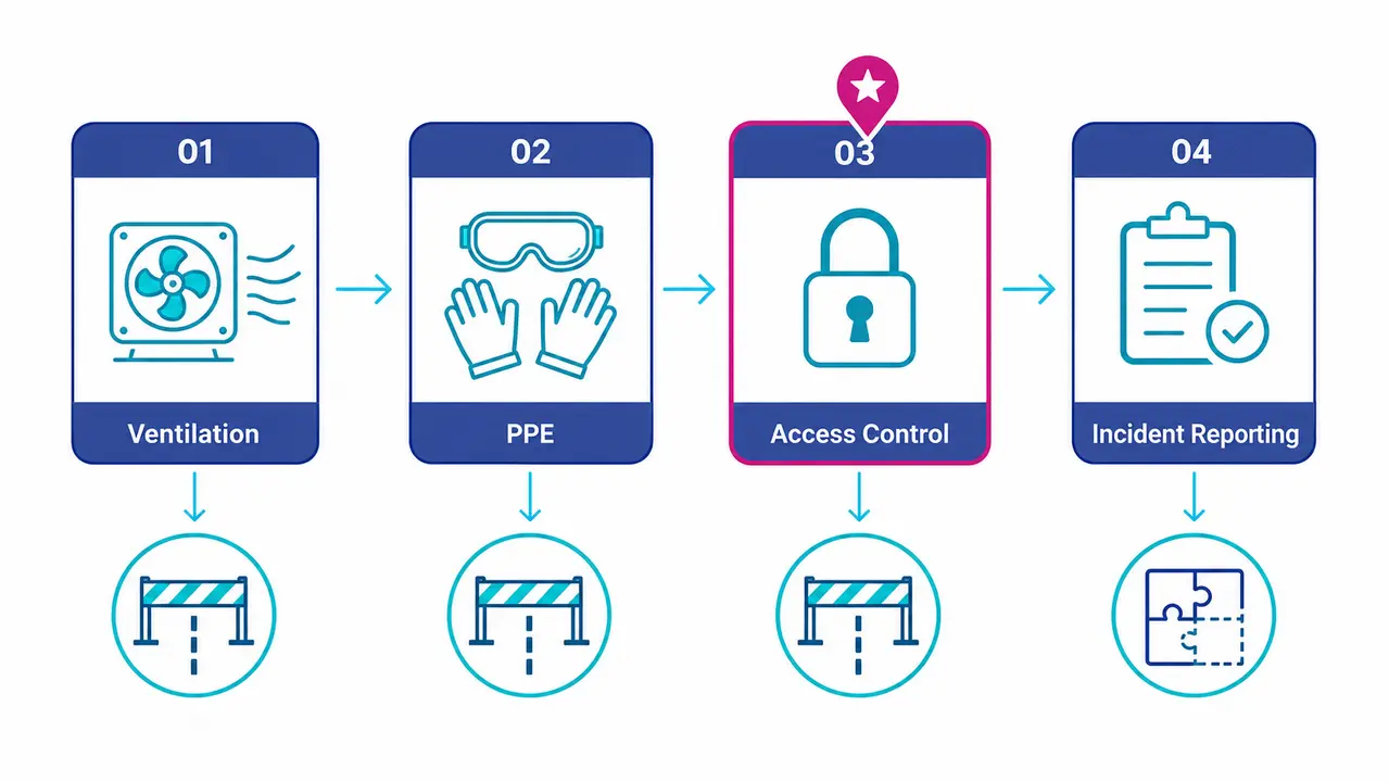
Programs must implement formal safety governance covering ventilation, chemical exposure controls, personal protective equipment, training and written procedures aligned with institutional environmental health requirements. Typical controls include ventilation engineering, exposure monitoring where applicable, restricted access, incident reporting and competency-based onboarding for learners.
Occupational formaldehyde exposure is a specific regulatory concern. The Occupational Safety and Health Administration maintains strict standards for formaldehyde exposure in laboratories. Our facility adheres to these safety governance principles. Environmental and safety officers guarantee best practice according to guidelines.
Safety is not a static checklist. It is a continuous operational requirement enforced by dedicated officers within the facility.
Structuring a safety briefing for visiting faculty and CME learners
Visiting faculty and CME learners require a standardized safety briefing before entering the lab. The core components include conduct expectations, personal protective equipment requirements, prohibited activities, emergency procedures and documentation of training completion. Cadavers for medical students in CME contexts demand the same rigor as foundational courses.
Ensuring respectful practice and professional conduct
Respect is operationalized through clear codes of conduct, controlled access, appropriate language, privacy protections and explicit policies on photography and data use. Donor-centered language, restricted recording and formal reflection components are standard professional norms.
Our facility maintains a sterile, tidy and respected environment. A professional team handles the whole process. This is our operational proof of the ethical position. The National Library of Medicine publishes research on teaching professionalism using dissection-based anatomy, highlighting how explicit conduct policies reinforce the donor as a first patient.
An anatomy curriculum must codify these norms. Cadavers for medical students cannot be utilized without a binding professionalism framework that governs learner behavior.
Donor information and documentation for educational use
Availability of donor information varies by program and consent scope. It may include de-identified donor profiles and relevant medical history elements when permitted and necessary for the educational objective. Our facility provides documented donor medical history where law and donation terms permit. This includes age, weight, BMI, clinical conditions, imaging, genetic and hereditary information and prior surgeries.
Privacy-by-design dictates minimum necessary information. Documentation supports course planning without implying guaranteed access. A medical school cadaver utilized for a cardiac procedure rehearsal requires prior surgical history. A human cadaver medical school specimen used for a first-year block requires less granular clinical data.
Course directors must specify their documentation needs early. We provide what the law and the donation terms allow, matched to the specific research or educational need.
Choosing whole-body donors versus regional donors
Whole-body donors support integrated regional-to-system learning and longitudinal curricula. Regional donors efficiently support targeted procedural objectives and specialty courses. A first-year integrated anatomy block requires a whole-body donor. A focused workshop on extremity, head and neck, or thorax procedures can use a regional specimen.
Course-design examples dictate the choice. A cadaver lab medical school session focused on spinal anatomy requires a whole-body donor to trace nerve pathways. An anatomy curriculum elective focused on foot and ankle procedures requires only an extremity specimen. Alignment to outcomes, staffing and time-on-task determines the most efficient use of the resource.
Selecting a regional donor when a whole body is required limits the educational scope. Selecting a whole body when a regional donor suffices wastes biological material.
Assessing learning outcomes from cadaver-based anatomy
Assessment should combine objective anatomy and imaging checks with structured procedural checklists when applicable and professionalism criteria tied to lab conduct. Measurable outcomes include identification accuracy, imaging correlation performance, structured OSCE-style stations and faculty-rated professionalism behaviors.
Documentation is required for accreditation and continuous improvement. Cadavers for medical students yield data that must be captured. Our facility provides the environment and the equipment, including C-arm fluoroscopy and laparoscopic towers, to support these assessment activities. The course director owns the assessment framework and the resulting data.
Without structured assessment, a lab session is merely an exposure. Competence requires measurement.
Supporting medical device R&D and pre-clinical evaluation
With appropriate governance, consent alignment and scheduling controls, cadaver-based environments support device usability evaluation, procedural workflow testing and training content validation. R&D and clinical affairs leads require protocol-driven sessions, documentation standards, imaging support and a strict separation of educational versus R&D objectives.
A human cadaver medical school specimen used for device testing requires explicit consent for that purpose. Our facility supports this work by providing fully equipped surgery rooms with advanced monitoring and imaging equipment. We provide the human model, the imaging and the staffing. The R&D team provides the protocol and the device.
A cadaver lab medical school environment can serve dual purposes, but never simultaneously on the same specimen without explicit authorization. The separation of objectives is a governance requirement, not a logistical preference.
Planning a cadaveric course or pre-clinical study?
Tell us your educational or research objectives and we will match the right specimen, imaging and facility setup.
Frequently asked questions
Which preservation method suits a first-year anatomy block versus a procedural skills session?
Embalmed donors support longer-term anatomy teaching, where the same specimen is used across many sessions. Fresh-frozen or lightly preserved tissue is required where realistic handling matters, because a heavily embalmed specimen will not yield realistic tissue feedback for a laparoscopic tower exercise. Selecting the wrong modality invalidates the session, so the choice follows the learning objective rather than availability.
When does a course genuinely need a whole-body donor rather than a regional specimen?
A whole-body donor is needed when the objective requires tracing structures across regions. A session on spinal anatomy needs one to follow nerve pathways along their full course. A focused elective on foot and ankle procedures needs only an extremity specimen. Matching the specimen to the outcome avoids using a whole-body donor where a regional one would serve.
What can a cadaver lab assess that a simulator cannot?
Anatomical variation and tissue plane behaviour. A synthetic model reproduces surgical geometry but not the variability between individuals, so it cannot validate a step whose difficulty comes from that variability. Simulation remains stronger for repetition, standardisation and scalable assessment, which is why blended designs usually outperform either method alone.
How much donor medical history can a course director expect?
Availability varies with the consent scope of the donation programme. It may include a de-identified donor profile and the clinical history elements relevant to the stated objective. Privacy-by-design means the minimum necessary is released, so a cardiac procedure rehearsal may warrant prior surgical history while a first-year block does not.
What formaldehyde controls does a teaching laboratory need?
Occupational formaldehyde exposure is a specific regulatory concern with defined limits. Controls combine ventilation engineering, exposure monitoring where indicated, personal protective equipment, documented training and written procedures. These sit under formal safety governance rather than being left to individual practice.
Cadaveric education and pre-clinical evaluation depend on matching the right specimen, preservation method and governance framework to a defined objective. Our facility provides the human model, the imaging and the staffing under Ministry of Health supervision. Course directors and R&D teams provide the curriculum and the protocol. Contact our laboratory to discuss your next programme.
About Biotech Anatomy

Biotech Anatomy LTD has provided practical anatomy and surgical education from Science Park, Rehovot since 2017. We are the only private Israeli company dedicated to employing human cadaveric material for research, and we operate fully equipped surgery rooms and classrooms with advanced monitoring, imaging and surgical equipment. We support medical teams, academic researchers and medical device developers through the whole study, donor recruitment and matching to the research need, pre-scan imaging, import and preparation of the specimen, trained staff throughout the session, and proper treatment of the remains afterwards, all under Ministry of Health supervision. Specimens are imported fresh-frozen after procurement, and donor information and diagnostic data are made available as far as law and the terms of donation permit. Our facility exists to honour what body donors intended their donation to achieve.